We know that biological age and chronological age of the brain hardly coincide: the first indicates the age that the organ demonstrates based on its state, the second coincides with the years of life of its owner. Another thing we know is that a biologically older brain is more at risk for neurodegenerative diseases such as Alzheimer’s.
What we didn’t know, however, is that not all regions of the brain age in the same way: until now, magnetic resonance imaging calculated an overall age, a number that was supposed to tell us whether at fifty we had the brain of a twenty-year-old or an eighty-year-old; a study published in PNAS conducted a more detailed analysis of each area of the brain, finding not only that different brain regions age at different rates, but also which ones tend to age sooner.
A spatial map of local brain age
To reach their conclusions, the scientists trained an AI model on almost 15,000 MRI scans of the brains of as many healthy adults between the ages of 19 and 100. After training, the researchers tested the system on a group of nearly 2,000 people – 1,102 with normal cognitive functions, 354 with mild cognitive problems and 529 with Alzheimer’s. The result is what Andrei Irimia, one of the authors, calls “a spatial map of local brain age.”
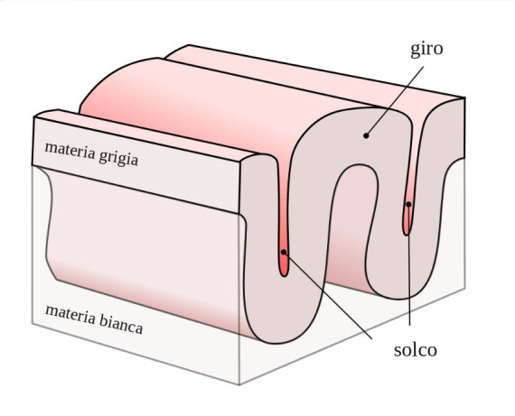

In cognitively healthy adults, the model’s estimates revealed a clear pattern: the frontal and temporal lobes, involved in complex thinking and memory, showed an older biological age than posterior regions, such as the visual processing centers of the occipital lobe (i.e. the areas of the visual cortex that process information arriving at the eyes). Even the surface of the cortex showed different aging rhythms: the grooves, i.e. the deep depressions between one fold and another, were older than the turns, the raised parts (see image).
Right or left: which side ages sooner?
There is also a broader trend that emerges from MRIs, namely that the right hemisphere tends to be older than the left. «The left-right asymmetry is particularly interesting», comments Irimia a Psypost: «we don’t yet know the cause, but it raises a question spontaneously (still unanswered, ed.) – do the two hemispheres age at different rates?”.
As for Alzheimer’s patients, as expected, MRI scans revealed more pronounced aging in the regions of the brain normally most affected by neurodegeneration, such as the pallidum, the putamen (which coordinate movements and some types of learning) and the hippocampus, which manages emotions and memory.
It’s just the beginning
The study completes a small corner of a gigantic puzzle: to find new pieces to join, it will be necessary to conduct new studies by following the participants for several years, as well as training the AI models on Alzheimer’s patients. Only in this way will we be able to understand whether the aging differences of the various brain regions can predict future brain health: at the moment, Irimia underlines, we cannot draw hasty conclusions based on brain scans lump sumbecause “an ‘older’ local brain age does not, in itself, mean that a person has Alzheimer’s or will develop it.”