Centering is an essential organelle in the cell, which plays a crucial role in various cellular processes. At present, both their function and structure are being studied and research in the field of cell biology. The centering is mainly composed of two centrioles, which are cylindrical structures located in the center of the centering and oriented perpendicularly with each other. The main function of the centers is the organization of microtubules and the promotion of tubulin polymerization, which allows the formation of mitotic spindle during cell division.
In this ecology article, we will learn the Center function and its structure. Likewise, you can know the cycle through which the centers go through, differentiate between a centering and centriole, and more.
What is the centers
The centers, also known as Citocenter, is a Structure close to the cell nucleus that plays a crucial role in cell division. In animal cells, the centers consists of two centrioles, which are structures composed of microtubules. These centrioles are of great importance, since they contribute to the formation of mitotic spindle, which is necessary for cytokinesis.
Although centersoms do not have their own membranes, they are associated with nuclear envelope. It is important to highlight that the two centrioles that make up the centering are paired and immersed in a set of protein called “pericentriolar material”. This pericentriolar material is an optically dense material.

Center function
Main functions:
- Organization of microtubules: The centers plays a fundamental role as the “organizing center of the microtubules”. Its main function is to organize and promote the polymerization of tubulin, the main protein of microtubules.
- Segregation of chromosomes: During the cell division, the centers participate in the formation of the mitotic spindle, which connects the chromosomes with the poles of the cell. This is essential to achieve equitable segregation of chromosomes in daughter cells.
Secondary functions:
- Maintenance of cell form: Centersoms play a role in maintaining cell form.
- Cellular movements: They participate in the movements of the membranes, since they are related to the microtubules and other components of the cytoskeleton.
- Genome stability: Recent studies suggest that centers are also involved in genome stability.
Center cycle
Phase G1
During the G1 phase of the cell cycle, Each cell contains a single centering. However, when the cell enters the S phase and the replication of DNA begins, the replication of the centering also occurs. In this phase the centrioles of the centering lose their orthogonal disposition.
Phase s
At the beginning of the S phase, the Duplication of the centriolesboth of the mother and centriolo of the centriole son. Structures called proceedings are formed. At the end of the S phase, the elongation of the processes occurs.
G2 phase
During the G2 phase, there is a Separation of the two original centrioles and their respective proceedings in training. This separation results in the distribution of pericentriolar material, resulting in the formation of two centers. In the transition between phase G2 and phase M, there is an important change in the centers known as the maturation of the centering. Before mitosis, centrioles begin to recruit more pericentriolar material; For example, an increase in the number of γ-tubulin rings, which increases its ability to generate microtubules.
Phase m
During phase M, the centersoms play a fundamental role in the Mitotic spindle formation. Microtubules are formed from the pericentrione matrix of each center. The cell division plane by which the stem cell is divided into two is always perpendicular to the axis of the mitotic spindle and is generally equidistant between the two centers.

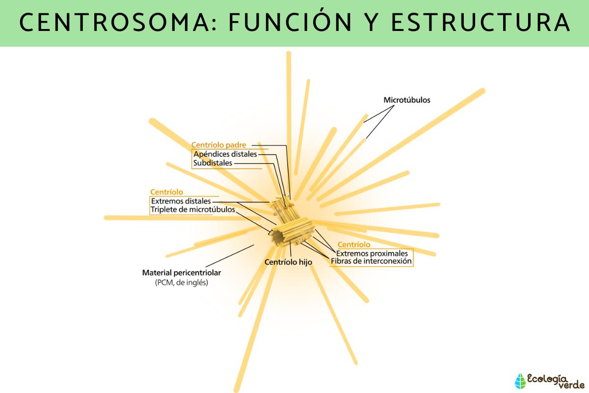
Center structure
The Center and Centriolo are cellular components that can often be confusing, however, based on the concepts that we will develop below it will be easier for us to recognize and differentiate them.
A Centering consists of two centrioles or diplosome (2 centrioles) and is located in the center of the center. The structure of the centrioles is similar to that of the basal corpuscles of the cilia. The centrioles are oriented perpendicularly to each other and have a cylinder shape, with walls composed of nine microtubules triplets, without a central microtubule, forming the structure called 9+0. Microtubules triplets are united by a Nexin protein bridge.
The microtubules in the centriole are divided into three types:
- Microtubule A (internal): It presents a circular section (13 protofilaments) and is closer to the cylinder axis.
- Microtubule B: It is located between microtubules A and C. Its section has a crescent shape and shares three protofilaments with microtubule A.
- Microtubule C (external): It has a medium -shaped section and shares three protofilaments with microtubulum B.
The set of centrioles and pericentriolar material is called MicroTubules Organizing Center (COMT). Another relevant structure is The rsterwhich consists of fibers formed by microtubules that grow and organize in a radial pattern around the centering. The microtubules of the rster give rise to the microtubules of the acromatic spindle during cell division.
Now that you know the function of the centering and its structure, you may also be interested in these other articles on the plasma membrane: what is, functions and structure and the smooth endoplasmic reticulum: what is and function.
If you want to read more articles similar to CENTROOMA: FUNCTION AND STRUCTUREwe recommend that you enter our biology category.
- Bornens M. (2012). The Centers in Cells and Organisms. Science. 335: 422-426.
- Fu j, do im, glover dm. (2015). The centers and its duplication cycle. Cold Spring Harbor Perspectives in Biology. 7: A015800.